Home    中文  
 
  • Search
  • lucene Search
  • Citation
  • Fig/Tab
  • Adv Search
Just Accepted  |  Current Issue  |  Archive  |  Featured Articles  |  Most Read  |  Most Download  |  Most Cited
Clinical Research

Evaluation of therapeutic effects for drug induced liver injury and viral hepatitis using quantitative ARFI technology

  • Jiyuan Ren 1 ,
  • Zhimin Liu 1 ,
  • Chunxing Li 1 ,
  • Xuechun Xu 1 ,
  • Ruoling Han , 2,
Expand
  • 1.Department of Ultrasound, the No. 2 Hospital of Baoding, Baoding 071051, China
  • 2.Department of Ultrasound, the Fourth Hospital of Hebei Medical University, Shijiazhuang 050011, China
Corresponding author: Han Ruoling, Email:

Received date: 2021-06-02

  Online published: 2022-06-21

Copyright

Copyright by Chinese Medical Association No content published by the journals of Chinese Medical Association may be reproduced or abridged without authorization. Please do not use or copy the layout and design of the journals without permission. All articles published represent the opinions of the authors, and do not reflect the official policy of the Chinese Medical Association or the Editorial Board, unless this is clearly specified.

Abstract

Objective

To explore the value of acoustic radiation force pulse (ARFI) imaging in the evaluation of therapeutic effects for drug induced liver injury (DILI) and viral hepatitis (VH).

Methods

Seventy-four inpatients diagnosed with DILI or VH at Department of Gastroenterology of the No. 2 Hospital of Baoding from January 2017 to August 2020 who met the inclusion criteria were included in this study. Conventional ultrasound and ARFI examinations were performed for each patient before treatment, one month after treatment, and three months after treatment, and virtual touch tissue quantification (VTQ) values were acquired and recorded. One hundred healthy volunteers were included as controls and VTQ values of the liver were also acquired and recorded.

Results

There were 34 cases of DLIL and 40 cases of VH included in this study. There was no significant difference in VTQ values between the DILI group and HV group before treatment (1.74±0.18 m/s vs 1.70±0.41 m/s, P>0.05), while they were both significantly higher than those of normal volunteers (1.12±0.16 m/s, P<0.05). There was no significant difference in VTQ values between one month and three months after treatment in the DILI group (1.14±0.23 m/s vs 1.06±0.40 m/s, P>0.05), while they were both significantly lower than those before treatment (P<0.05). There was no significant difference in VTQ values between before treatment and one month after treatment (1.59±0.16 m/s) in the HV group (P>0.05), while they were both significantly higher than those three months after treatment (1.12±0.23 m/s, P<0.05). There was a significant difference in VTQ values between DILI group and HV group one month after treatment (P>0.05).

Conclusion

As a non-invasive and quantitative method, ARFI can assess liver stiffness objectively and observe the stiffness changes in DILI and HV after treatment dynamically. It may provide information about the cause of liver injury according to the patients' recovery cycle.

Cite this article

Jiyuan Ren , Zhimin Liu , Chunxing Li , Xuechun Xu , Ruoling Han . Evaluation of therapeutic effects for drug induced liver injury and viral hepatitis using quantitative ARFI technology[J]. Chinese Journal of Clinicians(Electronic Edition), 2022 , 16(03) : 264 -267 . DOI: 10.3877/cma.j.issn.1674-0785.2022.03.013

声辐射力脉冲(acoustic radiation force impulse,ARFI)成像技术是一种新型、无创、定量的超声弹性成像技术。它主要利用超声探头发射推力脉冲波,在组织内形成一个推力,通过测量组织在推力作用下产生的剪切波速度,来反映组织的弹性信息。ARFI成像技术是近几年超声领域研究的热点。研究表明,ARFI在评估肝脏纤维化、急性肝损伤、脂肪肝等方面有重要的临床意义1, 2, 3。但是,ARFI技术对于肝损伤治疗效果评价的价值,相关研究尚不多见。本研究旨在探讨ARFI技术对药物性肝损伤(drug induced liver injury,DILI)与病毒性肝炎(viral hepatitis,VH)治疗效果评价的价值。

资料与方法

一、对象

选择2017年1月至2020年8月来河北省保定市第二医院消化科就诊并住院治疗诊断为DILI或VH患者为研究对象。入组标准:(1)有明确的病理诊断结果;(2)治疗前及治疗后1、3个月均行常规超声检查及ARFI检查;(3)治疗后3个月内患者临床症状缓解,肝功能恢复正常水平;(4)临床资料完整且愿意参加本临床研究并签署知情同意书。排除标准:(1)患者呼吸不能配合,ARFI检查不成功;(2)未完成标准治疗疗程。治疗方法参照《慢性乙型肝炎防治指南(2015更新版)》4及《药物性肝损伤诊治指南》5。同时设对照组,选取同期在本院消化科门诊就诊且肝功能无异常,常规超声检查肝脏无明显弥漫性病变及局灶性病变的志愿者100名。本研究经本院伦理委员会批准,所有受检者均签署知情同意书。

二、仪器与方法

采用西门子S3000彩色多普勒超声诊断仪,腹部凸阵探头6C1 HD,探头中心频率3.5 MHz。所有患者超声检查前需空腹8 h以上。患者平卧位或左侧卧位于检查床上,先行常规二维超声及彩色多普勒扫查肝脏,观察肝脏的形态、大小、内部回声、肝内血流分布以及是否有局灶性病变等。嘱患者保持平静呼吸,探头垂直放置于右侧肋间隙显示肝右前叶。保持探头位置固定不动,启动ARFI成像模式,感兴趣区放置深度为3.5~5.5 cm,避开肝内管道结构,嘱患者暂停呼吸至少5 s,按update键,读取并记录所得的声触诊组织定量(virtual touch tissue quantification,VTQ)值及取样深度。同一位置重复测量5次,取平均值记为VTQ值。
对照组超声检查前需空腹8 h以上。先行常规超声检查排除肝脏弥漫性病变及肝局灶性病变。随后启动ARFI模式,检查方法及数据收集同肝炎患者。

三、统计学分析

应用SPSS 23.0统计软件进行数据分析,计量资料以
x¯
±s表示,多组间比较采用单因素方差分析,组间两两比较采用最小显著性差异法(least significant difference,LSD);2组间比较采用t检验。以P<0.05为差异有统计学意义。

结果

一、患者基本资料

本研究DILI组患者34例,其中,男性14例,女性20例;年龄20~59岁,平均(32.3±2.9)岁。入组VH患者40例,其中,男性24例,女性16例;年龄33~68岁,平均(44.6±3.3)岁。入组志愿者100名,其中,男性58名,女性42名;年龄29~63岁,平均(38.4±2.5)岁。治疗后1个月DILI组患者各肝功能指标均较治疗前显著降低(P<0.05,表1),治疗后3个月VH组患者各肝功能指标均较治疗前显著降低(P<0.05,表2)。
表1 药物性肝损伤患者治疗前后肝功能变化情况(
x¯
±s
时间 AST ALT TBIL DBIL
治疗前 217.78±41.42 289.64±38.45 135.38±50.32 108.56±55.28
治疗后1个月 97.60±30.31a 114.32±29.81a 78.67±32.81a 63.92±31.08a
治疗后3个月 47.72±24.41ab 53.61±23.40ab 40.78±27.68ab 29.70±17.80ab
F 241.313 525.719 52.852 36.763
P <0.001 <0.001 <0.001 <0.001

注:与治疗前比较,aP<0.05;与治疗后1个月比较,bP<0.05;AST为谷草转氨酶;ALT为谷丙转氨酶;TBIL为总胆红素;DBIL为直接胆红素

表2 急性病毒性肝炎患者治疗前后肝功能变化情况(
x¯
±s
时间 AST ALT TBIL DBIL
治疗前 249.58±37.38 379.27±50.36 74.90±32.79 61.31±30.78
治疗后1个月 130.51±30.03a 215.98±34.76a 66.37±29.86 56.20±28.12
治疗后3个月 69.74±20.96ab 92.36±21.23ab 52.41±20.96a 41.07±23.42ab
F 366.735 592.419 6.429 5.814
P <0.001 <0.001 0.002 0.004

注:与治疗前比较,aP<0.05;与治疗后1个月比较,bP<0.05;AST为谷草转氨酶;ALT为谷丙转氨酶;TBIL为总胆红素;DBIL为直接胆红素

二、肝炎患者治疗前后VTQ值比较

单因素方差分析显示,治疗前DILI组、治疗前VH组及对照组间肝脏VTQ值差异有统计学意义(P<0.05);组间两两比较显示,治疗前DILI组与VH组肝脏VTQ值差异无统计学意义(P>0.05),且2组肝脏VTQ值均高于对照组[(1.12±0.16)m/s],差异有统计学意义(P<0.05,表3)。
表3 2组受试者治疗前后声触诊组织定量值比较(m/s,
x¯
±s
组别 例数 治疗前 治疗1个月 治疗3个月 F P
DILI组 34 1.74±0.18 1.14±0.23a 1.06±0.40 a 57.438 0.000
VH组 40 1.70±0.41 1.59±0.16 1.12±0.23ab 46.180 0.000
t 0.527 9.882 0.805
P 0.600 <0.001 0.423

注:与治疗前比较,aP<0.05;与治疗后1个月比较,bP<0.05

DILI组治疗前及治疗后1、3个月的肝脏VTQ值差异有统计学意义(P<0.05);组间两两比较结果显示,治疗后1、3个月肝脏VTQ值差异无统计学意义(P>0.05),两者均显著低于治疗前,差异有统计学意义(P<0.05,图1表3)。
图1 药物性肝损伤患者治疗前后ARFI成像图。图a为治疗前,声触诊组织定量(VTQ)值为1.48 m/s;图b为治疗后1个月,VTQ值下降为1.15 m/s;图c为治疗后3个月,VTQ值为1.09 m/s
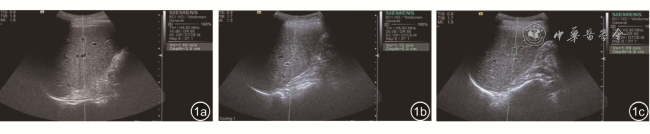
VH治疗前组及治疗后1、3个月的肝脏VTQ值差异有统计学意义(P<0.05);组间两两比较结果显示,治疗前与治疗后1个月的肝脏VTQ值差异无统计学意义(P>0.05),两者均显著高于治疗后3个月,差异有统计学意义(P<0.05,表3)。
治疗后1个月VH组肝脏VTQ值显著高于DILI组,差异有统计学意义(P<0.05);治疗前及治疗后3个月2组间肝脏VTQ值差异无统计学意义(P>0.05,表3)。

讨论

DILI是消化内科常见的肝脏疾病之一,由药物自身或其代谢物诱发,是最常见且严重的药物不良反应之一,以发病急、进展快、病死率高为特征,其发病率占急性肝损伤半数以上,至今仍缺乏简便、客观、特异的诊断指标6。VH是世界范围内最重大的公共卫生安全问题之一,具有发病率高、高度遗传易感性的特点,威胁性较高7
肝纤维化、肝硬化是许多慢性弥漫性肝病的病理结局。肝脏纤维化是一个动态的发展过程,因此早期确诊并及时治疗可以减缓其发展进程。肝脏穿刺活检是弥漫性肝病的诊断金标准,但因其有创性而存在许多局限性。ARFI成像技术是一种安全无创、简便快捷、可重复性好的成像方法,对肝纤维化的分级与评估具有重要意义8, 9。本研究结果显示,与对照组相比,DILI组与VH组治疗前肝脏VTQ值明显增高,提示两种不同病因所致的肝脏病变,均在疾病发展过程中导致肝脏硬度增加,与文献10, 11报道的研究结果一致。
本研究结果表明,DILI组治疗后1个月肝脏VTQ值较治疗前明显下降,与治疗后3个月肝脏VTQ值无显著差异,提示DILI经过有效治疗后,肝脏硬度会较快恢复正常水平。而VH治疗后,1个月肝脏VTQ值较治疗前略下降,显著高于治疗后3个月肝脏VTQ值,提示VH经过有效治疗后,肝脏硬度恢复较慢,但在治疗后3个月会恢复至正常水平。分析原因可能DILI是急性损伤作用,解除致病因素以后,肝细胞恢复快,转氨酶下降明显,而VH是慢性病毒的损伤过程,肝细胞一直受病毒的攻击,尽管致病因素缓解,但还存在一定的致病因素,肝细胞恢复慢。
本研究发现,DILI组治疗后1个月肝脏VTQ值显著低于VH组,进一步提示VH恢复周期长,DILI恢复周期较短。对于部分病因诊断困难的肝损伤患者,如果治疗1个月患者肝脏VTQ值下降明显,倾向于诊断DILI,如果VTQ值下降不明显,倾向于诊断VH。
本研究具有一定的局限性。首先,本研究病例数较少,结果尚需要加大样本量进一步验证。其次,本研究中ARFI结果未与临床肝功能等指标进行比较。
综上所述,ARFI技术作为一种无创定量的超声弹性成像技术,可以客观评价肝组织硬度及其变化,动态观察DILI及VH治疗前后肝脏的硬度变化,并可根据患者的恢复周期为临床肝炎的病因诊断提供依据。
1
Dietrich CF, Bamber J, Berzigotti A, et al. EFSUMB guidelines and recommendations on the clinical use of liver ultrasound elastography, update 2017 (Long Version) [J]. Ultraschall Med, 2017, 38(4): e16-e47.

2
Bâldea V, Sporea I, Tudor A, et al. Virtual touch quantification using acoustic radiation force impulse imaging technology versus transient elastography for the noninvasive assessment of liver fibrosis in patients with chronic hepatitis B or C using liver biopsy as the gold standard [J]. J Gastrointestin Liver Dis, 2020, 29(2): 181-190.

3
涂春华, 李嘉, 王春妍, 等. 超声内镜、Fibroscan、声辐射力脉冲成像、血清学指数及其联合对早期肝硬化的诊断价值 [J]. 中华肝脏病杂志, 2019, 27(8): 615-620.

4
王贵强, 王福生, 成军, 等. 慢性乙型肝炎防治指南(2015更新版) [J]. 中华肝脏病杂志, 2015, 8(6): 888-905.

5
于乐成, 茅益民, 陈成伟. 药物性肝损伤诊治指南 [J]. 中华肝脏病杂志, 2015, 23(11): 810-820.

6
European Association for the Study of the Liver. EASL Clinical practice guidelines: drug-induced liver injury [J]. J Hepatol, 2019, 70(6): 1222-1261.

7
Charlton MR, Alam A, Shukla A, et al. An expert review on the use of tenofovir alafenamide for the treatment of chronic hepatitis B virus infection in Asia [J]. J Gastroenterol, 2020, 55(9): 811-823.

8
Ferraioli G, Wong VW, Castera L, et al. Liver ultrasound elastography: an update to the world federation for ultrasound in medicine and biology guidelines and recommendations [J]. Ultrasound Med Biol, 2018, 44(12): 2419-2440.

9
Ding H, Ma JJ, Wang WP, et al. Assessment of liver fibrosis: the relationship between point shear wave elastography and quantitative histological analysis [J]. J Gastroenterol Hepatol, 2015, 30(3): 553-558.

10
Cao W, Zhou Y, Niu Y, et al. Quantitative analysis of hepatic toxicity in rats induced by inhalable silica nanoparticles using acoustic radiation force imaging [J]. J Ultrasound Med, 2017, 36(9): 1829-1839.

11
Guo H, Liao M, Jin J, et al. How intrahepatic cholestasis affects liver stiffness in patients with chronic hepatitis B: a study of 1197 patients with liver biopsy [J]. Eur Radiol, 2020, 30(2): 1096-1104.

Outlines

/

京ICP 备07035254号-20
Copyright © Chinese Journal of Clinicians(Electronic Edition), All Rights Reserved.
Tel: 010-57830845 E-mail: zhlcyszz@cma.org.cn
Powered by Beijing Magtech Co. Ltd